


- clinic services
Comprehensive ultrasound diagnostics
Modern equipment enables accurate, comprehensive diagnostics of almost any part of the body.
Offer
Examination packages

Infant check-up package
3 scans for the price of 2
- transfontanelle head ultrasound
- abdominal cavity
- Graf method hip dysplasia screening
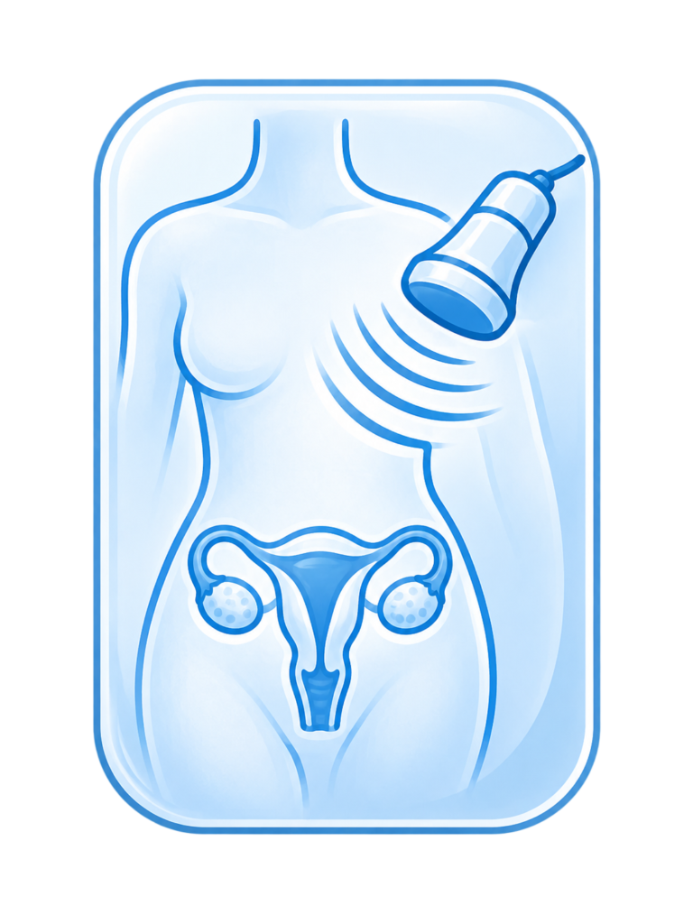
Women’s check-up package
3 scans for the price of 2
- thyroid
- breasts
- abdominal cavity
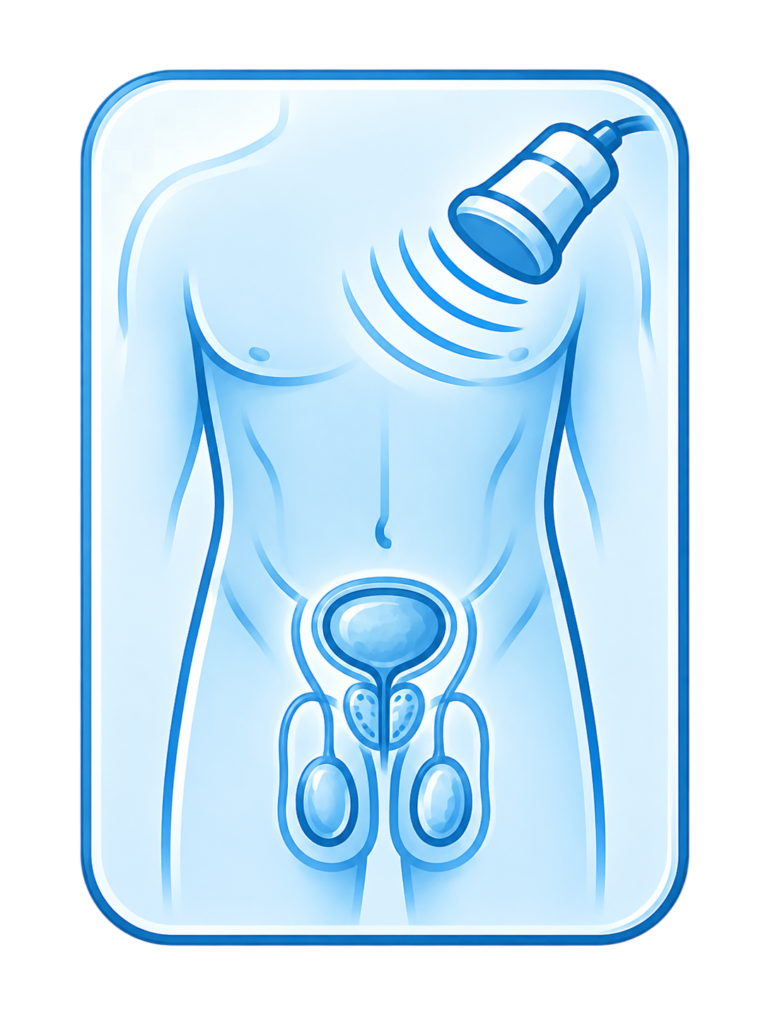
Men’s check-up package
3 scans for the price of 2
- thyroid
- abdominal cavity
- testicles
- Examinations
Internal medicine ultrasound
Internal medicine ultrasound enables a safe, non-invasive assessment of the internal organs, including the abdomen, thyroid, breasts, urinary tract and lymph nodes. The examination helps detect abnormalities, monitor previously diagnosed changes and determine whether further diagnostics are needed. The patient receives the results immediately after the examination.
Breast ultrasound allows for diagnostics of the breasts and nipples, assessment of their structure and imaging of the axillary lymph nodes. Ultrasound can detect different glandular changes, including malignant tumors. Alongside mammography, it is an important preventive examination and is recommended at least once a year. Breast ultrasound requires no special preparation. It is a non-invasive imaging test that enables a detailed breast assessment and helps identify various pathological changes.
Main indications for breast ultrasound
Breast diagnostics are usually performed to assess changes detected during a physical examination, mammography or other diagnostic tests. The examination can help identify pathological changes such as tumors, cysts or abnormalities in the blood vessels.
Benefits of breast ultrasound
Ultrasound offers many benefits in breast diagnostics. It is safe and non-invasive, with no exposure to harmful radiation. It is also highly accurate and allows detailed assessment of breast changes, especially in women with dense breast tissue. It can be used in both women and men.
Basic breast anatomy
The breast is a mammary gland located on the front wall of the chest. It consists of glandular tissue, fatty tissue and connective tissue. The breasts are supplied by the thoracic arteries, and secretions are carried through the milk ducts to the nipple.
Common breast conditions
The breasts may be affected by many diseases and disorders. Breast tumors are among the most common abnormalities detected by ultrasound. They may be benign or malignant and may require further diagnostics and treatment. Breast cysts are also common changes that can be assessed with ultrasound. Breast cancer is a serious condition that may require surgery, radiotherapy or chemotherapy.
Breast ultrasound is a safe and effective diagnostic examination that enables accurate breast assessment and detection of tumors, cysts and other changes. It supports early diagnosis, which increases the chance of effective treatment. If there are any symptoms or indications for the examination, it is always best to consult a specialist who will select the appropriate diagnostic methods and plan further care.
Abdominal ultrasound is one of the most common ultrasound examinations. It is worth performing preventively at least once a year. It allows the doctor to assess internal organs such as the liver, pancreas, spleen, kidneys and bladder, and to detect abnormalities in their structure. The examination can also help locate tumors, which may be important in cancer prevention. Abdominal ultrasound should be performed on an empty stomach. It is a non-invasive diagnostic test that uses advanced ultrasound technology to provide images of abdominal organs and identify abnormalities, pathological changes or structural defects.
Testicular ultrasound is performed in cases of testicular pain or enlargement of one or both testicles. It is also recommended when testicular pain occurs. Ultrasound allows assessment of the size and structure of the testicle, epididymis and scrotum. The examination requires no special preparation. If you have additional questions, please contact us by phone. Testicular ultrasound is a diagnostic procedure that assesses the structure, shape and function of the testicles in men. Using advanced ultrasound technology, it provides real-time images and helps identify pathological changes, injuries, cysts or tumors. During the examination, the ultrasound specialist moves the probe over the surface of the scrotum. This makes it possible to assess testicular structure and size, detect tumors, cysts or infiltrates, and evaluate blood flow in the vessels supplying the testicles. Ultrasound is particularly useful in diagnosing testicular inflammation, cysts, inguinal hernia and testicular cancer. It can also be used to monitor patients after testicular surgery or to assess treatment effectiveness.
Thyroid ultrasound is performed when palpable thyroid changes, enlarged neck circumference or abnormal TSH levels are present. The examination can detect thyroid abnormalities such as nodules and cysts. It requires no special preparation.
Thyroid ultrasound is a safe and non-invasive examination that uses sound waves to image the thyroid gland. During the examination, the patient lies on their back and gel is applied to the neck to improve image quality. The ultrasound probe is then used to obtain thyroid images on the monitor. The examination is painless and usually takes about 15 minutes. Thyroid ultrasound can help detect various thyroid conditions, including:
- Thyroid size: it helps determine thyroid size and detect enlargement or reduction.
- Thyroid tumors: it can detect thyroid tumors, including malignant tumors that may require further testing and treatment.
- Autoimmune diseases: it can detect autoimmune thyroid diseases such as Hashimoto disease and Graves-Basedow disease.
- Thyroid cysts: it can detect cysts that may be associated with other thyroid conditions.
- Treatment monitoring: it can also be used to monitor treatment of thyroid diseases such as Graves-Basedow disease, Hashimoto disease or thyroid cancer.
Thyroid ultrasound may also be used to diagnose and monitor thyroid disease in pregnant women, as these conditions can affect fetal development and maternal health.
Most common thyroid conditions
- Graves-Basedow disease: an autoimmune disease causing hyperthyroidism. Symptoms may include rapid pulse, hand tremor, rapid weight loss, sleep disorders and eye problems such as dryness and redness.
- Hashimoto disease: an autoimmune disease causing hypothyroidism. Symptoms may include fatigue, sleepiness, weight gain, dry hair and skin, and muscle and joint pain.
- Thyroid cancer: a malignant thyroid tumor. Common types include papillary, follicular and anaplastic thyroid cancer. Symptoms may include neck swelling, shortness of breath, hoarseness, swallowing difficulty and pain around the thyroid.
- Thyroid cysts: fluid-filled spaces in the thyroid. They may be associated with other thyroid conditions and often cause no symptoms, although in some cases they may cause pain, pressure or shortness of breath.
- Thyroid nodules: usually benign, but in some cases malignant. Symptoms depend on size and location and may include neck swelling, swallowing difficulty, shortness of breath and hoarseness.
Thyroid ultrasound is a safe, non-invasive examination that can help detect thyroid nodules, autoimmune diseases and thyroid cancer. Regular thyroid check-ups support early detection and prevention of complications. If you notice symptoms such as neck swelling, swallowing difficulty, shortness of breath or weight changes, consult a doctor for advice and further diagnostics.
Salivary gland ultrasound allows assessment of gland size and morphology. It can help diagnose tumors and gland enlargement. The examination is recommended when pain or swelling appears in the salivary gland area. It requires no special preparation. Salivary gland ultrasound is a non-invasive diagnostic test used to assess the structure, size and possible changes in the salivary glands. The parotid, submandibular and sublingual glands produce saliva, which plays an important role in digestion and oral health.
Neck ultrasound supports diagnostics of structures such as cervical lymph nodes, trachea, thyroid and salivary glands. It should be performed when the neck circumference increases or asymmetry appears. Enlarged cervical lymph nodes may also be an indication. The examination requires no special preparation. Neck ultrasound is a non-invasive diagnostic test that allows assessment of anatomical structures within the neck, including the thyroid, lymph nodes, trachea, carotid arteries, salivary glands, muscles and other tissues.
Cervical lymph node ultrasound allows assessment of the normal structure, size and morphology of lymph nodes in the neck. Enlarged cervical nodes often accompany a common cold, but they may also signal more serious conditions, including malignant diseases. The examination helps assess changes in lymph node size and determine whether a node is enlarging or has undergone fibrosis. During lymph node ultrasound, the specialist visualizes lymph nodes in different body areas, such as the neck, armpits, groin and abdomen, assessing their size, shape, symmetry and any abnormalities such as enlargement, structural changes or tumors.
Cervical lymph node ultrasound requires no special preparation.
Axillary lymph node ultrasound allows assessment of lymph node size, structure and morphology. It helps determine whether a node is enlarged or has undergone fibrosis. Lymph node ultrasound is recommended in cancer prevention, as enlarged lymph nodes, especially cervical nodes, may be concerning if they remain enlarged for more than two weeks. In such cases, biopsy is often performed in addition to ultrasound to provide a complete clinical picture. During the examination, the ultrasound specialist focuses on visualizing and assessing lymph nodes in the armpit area, evaluating their size, shape, symmetry and possible abnormalities such as enlargement, infiltrates or tumors.
Inguinal lymph node ultrasound allows assessment of lymph node size, structure and morphology. It helps determine whether a node is enlarged or has undergone fibrosis. Lymph node ultrasound is recommended in cancer prevention, as persistent enlargement may require further diagnostics, often including biopsy. Inguinal lymph node ultrasound assesses the structure, size, shape and possible abnormalities of lymph nodes in the groin area. These nodes play an important role in immunity and may be affected by different pathological processes.
Pleural cavity ultrasound helps diagnose many lung diseases and complications, including post-traumatic conditions. It is recommended for symptoms such as chronic cough, hemoptysis, chest pain, shortness of breath or breathing difficulties. The examination can help diagnose pneumonia, pleuritis, abscesses, embolism and increased fluid in the pleural cavity. It requires no special preparation. Pleural ultrasound is a diagnostic procedure used to assess the pleura, the serous membrane lining the inside of the chest and covering the lungs. It provides real-time images and helps identify pathologies, fluid accumulation or inflammatory changes.
Urinary tract ultrasound supports diagnostics of the bladder and prostate gland in men. Indications include, among others, blood in the urine. The examination can help diagnose tumors. Using an ultrasound probe, the specialist visualizes and assesses the urinary organs, including the kidneys, possible stones, tumors, cysts and other structural abnormalities. It also allows assessment of bladder volume and condition, as well as urine flow in the ureters.
Urinary tract ultrasound is performed in two stages. One hour before the examination, the patient should drink about one liter of water. The first stage is performed with a full bladder and the second after emptying it. The examination is performed on an empty stomach; smoking should also be avoided beforehand.
Subcutaneous lesion ultrasound is used to diagnose changes under the skin, such as lumps and thickened areas. Prevention and assessment of such changes is important, especially in cancer-related diagnostics, as subcutaneous lesions are often noticed accidentally by patients. During the examination, the ultrasound specialist visualizes the lesion and surrounding tissues to assess its structure, size, shape and possible abnormalities. The examination can also help distinguish benign lesions from potentially malignant ones.
Subcutaneous lesion ultrasound requires no special preparation.
Transfontanelle CNS ultrasound in infants allows assessment of brain tissue structures and fluid spaces within the skull. CNS ultrasound is a routine examination in premature babies. Indications also include low birth weight, low APGAR score, developmental disorders, increasing head circumference and abnormalities in a child’s neurological examination. The examination is performed through the anterior fontanelle.
It requires no special preparation.
Cranial suture ultrasound at our clinic in Lublin.
Pelvic ultrasound with assessment of the reproductive organs in patients under 16 at our clinic in Lublin.
- Examinations
Doppler ultrasound
It is based on a change in the frequency of a wave reflected from objects in motion. Blood cells in a vessel also move with the blood flow.
Doppler ultrasound is an ultrasound examination of blood vessels. It uses the Doppler effect, based on a change in the frequency of a wave reflected from moving objects. Blood cells in a vessel move with the blood flow. By comparing the emitted frequency with the reflected frequency, it is possible to calculate the speed and direction of blood flow. This is used to assess blood vessels for narrowing, dilatation and obstruction.
Doppler ultrasound is a fully safe and non-invasive diagnostic method that allows doctors to look inside the patient’s body. It is a precise tool that is indispensable in the diagnostics of many organs. It can detect many conditions at an early stage, making treatment more effective.
Doppler ultrasound of brain-supplying arteries allows assessment of blood flow in the vessels and helps estimate the degree of arterial narrowing. Doppler ultrasound observes the length of ultrasound waves reflected from blood vessels. This diagnostic procedure assesses blood flow in arteries supplying the brain, such as the internal carotid and vertebral arteries, which are essential for delivering oxygen and nutrients to the brain. The examination requires no special preparation.
Doppler ultrasound of carotid and vertebral arteries uses the Doppler effect to assess blood flow in arteries and veins. The method measures ultrasound waves flowing through and reflecting from blood vessels. It helps diagnose vascular narrowing. This diagnostic procedure assesses blood flow in the carotid arteries, which supply the brain, face, neck and other nearby structures.
Doppler ultrasound of the carotid and vertebral arteries requires no special preparation.
Doppler ultrasound of lower limb arteries is performed using the Doppler method, which observes ultrasound waves reflected from blood vessels. The examination is non-invasive and painless. It helps diagnose the location and degree of narrowing in lower limb arteries, which supply blood to the muscles, bones and tissues of the legs and support proper movement and function.
The examination requires no special preparation.
Doppler ultrasound of upper limb veins supports diagnostics of venous embolism and blood flow in the veins. The Doppler method is based on observing ultrasound waves. The examination is used in the diagnosis and treatment of venous thrombosis. During the examination, the ultrasound specialist directs ultrasound waves along the arms to visualize veins, assess blood flow, detect possible clots, and identify narrowing, dilatation or other venous abnormalities.
The examination requires no special preparation.
Doppler ultrasound of lower limb veins supports diagnostics of venous embolism and blood flow in the veins. The Doppler method is based on observing ultrasound waves. The examination is used in the diagnosis and treatment of venous thrombosis. During the examination, the ultrasound specialist directs ultrasound waves along the legs to visualize veins, assess blood flow, detect possible clots, and identify narrowing, dilatation or other venous abnormalities.
The examination requires no special preparation.
Aortic ultrasound is performed using the Doppler method, which monitors ultrasound waves. The examination is painless and non-invasive. Indications include back and spinal pain after degenerative disease has been ruled out, a diagnosed aneurysm, or a family history of aneurysm. The examination requires no special preparation. Aortic ultrasound is a non-invasive diagnostic test that assesses the anatomy and function of the aorta, the main artery of the body. It provides real-time images and helps identify abnormalities, pathological changes or structural defects.
- Examinations
Orthopedic ultrasound
Orthopedic ultrasound is a group of examinations used in orthopedics. It includes ultrasound of muscles, joints, tendons, bursae and other structures of the musculoskeletal system.
Muscle and tendon ultrasound is particularly useful for people exposed to injuries, for example due to increased physical activity such as athletes. The examination helps diagnose changes and abnormalities in muscle tissue.
Subcutaneous lesion ultrasound requires no special preparation.
Achilles tendon ultrasound helps diagnose structural abnormalities caused by injuries. Athletes are particularly prone to Achilles tendon injuries. Limited joint mobility is an indication for the examination. Achilles tendon ultrasound requires no special preparation. It is a diagnostic procedure that assesses the structure and condition of the tendon, the largest and one of the most important tendons in the human body, essential for walking, running and jumping.
Plantar fascia ultrasound allows assessment of fascial fibers and the size of swelling. The examination may be performed in the context of rheumatoid arthritis or systemic lupus erythematosus. Foot pain, for example during the first steps after getting out of bed in the morning, is an indication for the examination.
Knee joint ultrasound supports diagnostics of post-traumatic knee conditions and assessment of ligaments and tendons. It is performed when knee pain is present. The diagnostician assesses the joint cavity, kneecap, ligaments, tendons and menisci. Knee ultrasound requires no special preparation. It is a non-invasive and painless diagnostic test that provides detailed imaging of soft tissues and bones around the knee joint.
KNEE ANATOMY: The knee is a complex joint made of three bones: the femur, tibia and patella. It enables bending and straightening of the leg and includes many soft-tissue structures, such as tendons, ligaments, the joint capsule and cartilage, which provide stability and smooth movement.
HOW THE EXAMINATION IS PERFORMED: The patient lies on the examination table with the knee straight or slightly bent. Gel is applied to the skin to improve ultrasound transmission, and the doctor moves the ultrasound probe over the knee to obtain images of soft tissues and bones within the joint.
COMMON CONDITIONS: Knee ultrasound can detect various conditions, including:
- Bursitis: inflammation of the bursa around the knee, causing pain, swelling and movement difficulty. Ultrasound may show fluid in the bursa.
- Joint abscess: an inflammatory condition with pus accumulation inside the knee joint. Ultrasound may show fluid with calcifications and fibers.
- Bone fragments: loose bone fragments within the knee joint can cause pain and limited mobility and may be visible on ultrasound.
- Ligament injuries: ultrasound may show ligament damage associated with increased mobility and joint instability.
Graf method hip dysplasia screening ultrasound in infants enables early diagnosis of developmental hip dysplasia and helps assess its severity. The examination is usually performed at 5-6 weeks of age. It is carried out with the help of a parent, who holds the baby and helps maintain the correct position. Preluxation ultrasound is a diagnostic procedure used in infants to assess possible hip joint laxity, a condition in which the femoral head is not stably positioned in the acetabulum. It is important for early detection of hip development problems.
Hip joint ultrasound is performed to assess possible hip inflammation. During the examination, the bony outlines of the hip joint and fluid around the joint and greater trochanter are evaluated. Hip ultrasound is a diagnostic procedure that assesses the structure, function and possible changes in the hip joint, the largest and one of the most mobile joints in the human body.
Hip ultrasound requires no special preparation.
Hand joint ultrasound allows assessment of the structures of the carpal tunnels and fingers. It is performed in cases of hand pain, inflammation, sensory disturbances in the hand and fingers, and post-traumatic conditions. Hand joint ultrasound assesses the structure, condition and possible changes in the joints of the hand, including the wrist, metacarpus and phalanges.
Hand joint ultrasound requires no special preparation.
Forefoot ultrasound allows assessment of ligament structures, the joint capsule and forefoot tendons. It helps precisely diagnose causes of pain in the limb. The examination requires no special preparation. Forefoot ultrasound assesses the structure and possible changes in the forefoot area, including bones, joints, ligaments and muscles.
Pubic symphysis ultrasound in pregnant women at our clinic in Lublin.